


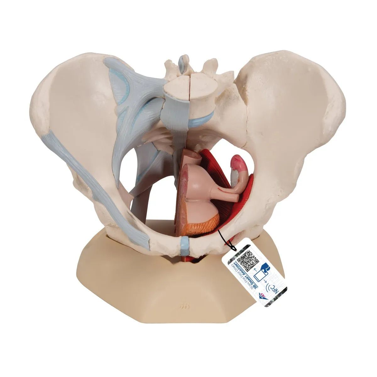

Female Pelvis with Ligaments Muscles and Organs - Includes 3B Smart Anatomy
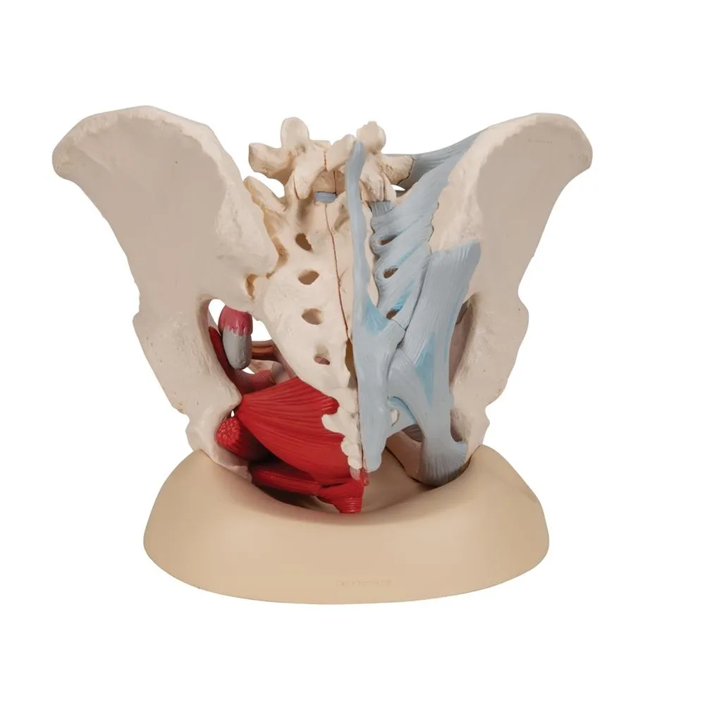
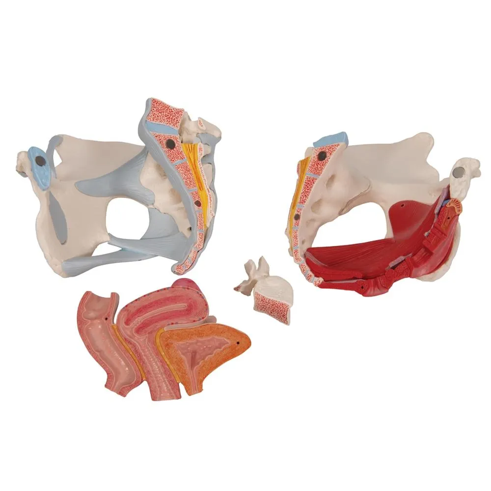
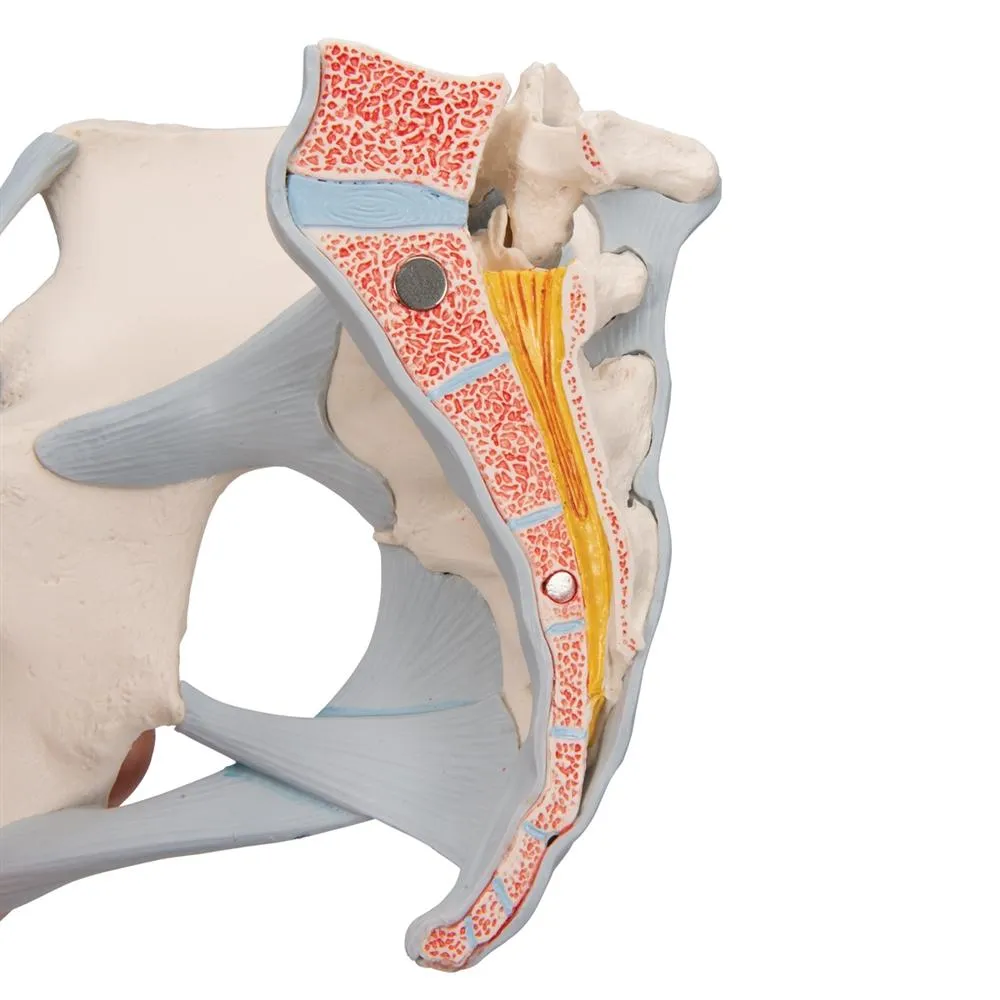
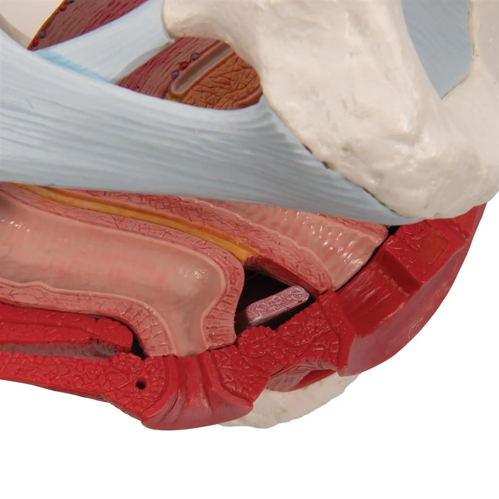
This life size four part model of a female pelvis represents detailed information about the topography of bones,
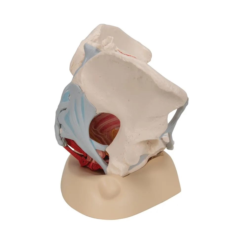
ligaments, pelvic floor muscles and female pelvic organs. The right half shows the bones with pelvic ligaments. In
addition, the left half of the pelvis contains the muscles of the pelvic floor including levator ani, ischiocavernosus,
deep and superficial transverse perineal, external anal sphincter, external urethral sphincter.
A partially removable bulbospongiosus demonstrates the vestibular bulb and Bartholin gland. The removable midsagital
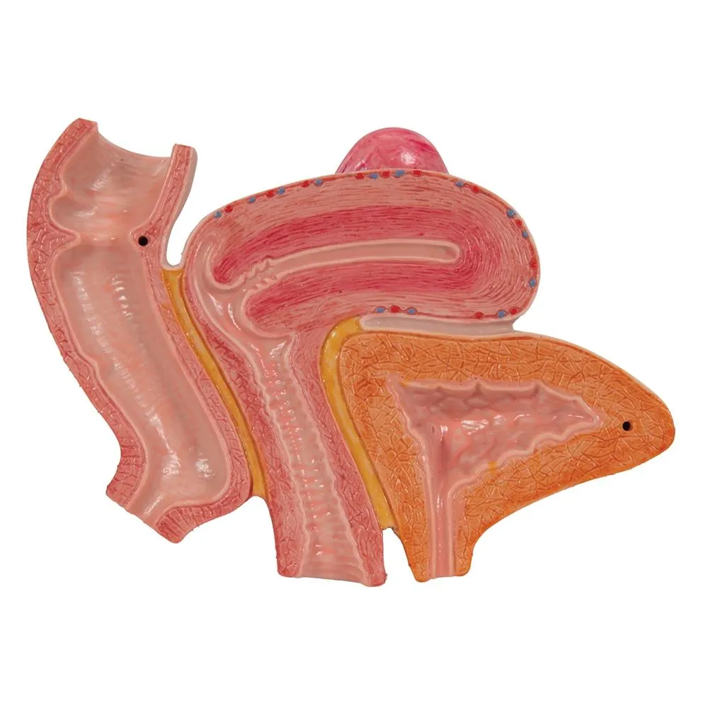
section through the urinary bladder, vagina, uterus and rectum demonstrates the relationship to the muscles of the
pelvic floor within its openings for urethra, vagina and rectum. The female genital organs are detailed for
gynecological and other anatomy studies.
GTSimulators by Global Technologies
3B Scientific Authorized Dealer.


 - Moss")


